
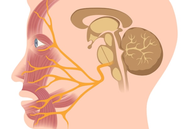
The Facial Nerve
The facial nerve is very important in Acoustic Neuroma surgery. We believe that facial nerve function outcome is the single most important factor in patient satisfaction after Acoustic Neuroma surgery. We cannot overemphasize the importance and priority to surgical strategies to preserve the facial nerve function during Acoustic Neuroma surgery and all treatment options take into consideration this highly important nerve.
The facial nerve runs together with the acoustic nerve in the skull base. This anatomical arrangement results in a close relationship between the facial nerve and the capsule of the Acoustic Neuroma which makes the nerve particularly vulnerable during surgery. As the tumor enlarges, the facial nerve is further stretched along the capsule of the tumor and consequently the risk to the nerve during tumor resection increases with the size of the tumor. Trauma to the facial nerve can cause temporary or permanent facial weakness with serious consequence. Surgery of Acoustic Neuroma requires maximal attention and priority to preservation of facial nerve function, particularly when treating large tumors. Facial nerve outcome is directly related to the expertise and experience of the skull base surgeon in Acoustic Neuroma surgery.
Innovations in Resecting Acoustic Neuroma and Preserving Facial Nerve Function:
Correctly identifying and preserving the facial nerve is critical during treatment of large Acoustic Neuroma. Specialized techniques to visualize and protect the facial nerve during Acoustic Neuroma surgery have been developed at The Center for Acoustic Neuroma.
When treating patients with large Acoustic Neuroma, we use an expanded translabyrinthine skull base approach in combination with an intra-capsular microsurgical tumor resection technique. The expanded translabyrinthine approach affords better visualization and control of the facial nerve throughout its course along the capsule of the tumor, allowing safer dissection of the tumor away from the facial nerve.
Over the years, careful observation of the anatomy of Acoustic Neuroma has led us to develop the concept of the intra-capsular tumor resection. This strategy is based on the concept that the neoplastic tissue in Acoustic Neuroma start centrally in the vestibulocochlear nerve (CN 8) and, as it grows, expands and thins out the nerve bundle, pushing the normal nerve fibers to the periphery of the tumor. Distinct attention to preserving the normal vestibular and cochlear nerve fibers during tumor resection provides an extra layer of protection to the facial nerve. In addition, at times, we may leave a thin layer of tumor over a portion of the facial nerve when the plane of dissection is poor between the tumor capsule and the facial nerve. This thin layer of tumor resembles a thin “carpet” of tumor that is adherent to the facial nerve and further attempts at removal of this slight tumor would put the facial nerve at grave risk. In our experience, this small residual tumor volume has not affected long term outcomes.
In summary, improvement of the surgical approach, refinement of the microsurgical technique for tumor resection, and long term experience and expertise have maximized the potential for favorable facial nerve outcomes even when treating very large Acoustic Neuroma.